Clinical History
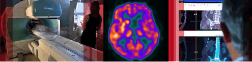
A 56 year old male smoker had a chest x-ray performed for investigation of a persistent cough. A suspicious mass was demonstrated in the left lower zone. A needle biopsy demonstrated non small cell lung carcinoma. The patient had no significant co-morbidities and surgical resection was considered as a treatment option. A PET (positron emission
tomography) scan was performed for staging.
Scan Findings
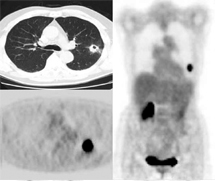
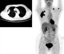
The PET scan (Figure One) demonstrates intense tracer uptake in the left lung mass and normal tracer uptake elsewhere. This indicates localised disease and the patient proceeded to surgery. A companion case is presented where the staging PET scan demonstrated metastatic disease (Figure Two). The presence of distant metastases precludes patients from curative surgical resection.
Discussion
The tracer used in PET is radio-labelled glucose which accumulates in areas of hypermetabolism. Many malignant neoplasms are hypermetabolic and PET imaging has demonstrated excellent utility in the diagnosis, staging and recurrence detection of many cancers. The main therapeutic approach for NSCLC is surgery for early disease as this provides the best chance of cure if the cancer hasn’t spread.
If the cancer has spread, surgery will not help the patient and PET is the most accurate test to determine if the cancer has spread. PET scans define local and distant disease better than any other modality and have a large impact on patient
management. A recent analysis of a database of over 30,000 Australian patients showed that staging of patients with several different cancer types changed in over 40% of patients following PET scanning.
Conclusion
PET improves the overall accuracy of clinical staging for NSCLC through better identification of nodal and distant metastases. By doing so, PET imaging has a significant impact on patient management by identifying those patients that will benefit most from surgical resection. It appears that PET will play a similarly powerful role in many other
malignancies.
Case Study submitted by
Dr Scott Beuzeville
Department of Nuclear Medicine,
St. George Hospital.
(PDF DOWNLOAD)
Figure 1. CT scan (left upper panel) and corresponding PET scan image (left lower panel) demonstrating avid tracer uptake in the lung cancer. Whole body PET scan (right panel) shows no uptake elsewhere to suggest metastatic disease. Right renal and bladder activity is physiologic.
Figure 2.Companion case demonstrating a pleural based right lung carcinoma (CT scan) and whole body PET scan. The PET scan
demonstrates uptake in the primary tumour (right lung) and several metastases (right sided ribs, left clavicle and the left humeral head). Physiologic cardiac, renal and bladder
activity is present.