Clinical History
A 75 year
old male suffered a mechanical fall at home. He was initially pain free but
developed progressive right hip pain over the following two days. Despite normal
radiographs, he was unable to weight bear and had difficulty caring for himself
at home. He was admitted to hospital for further management. On examination,
there was no shortening or rotation of the right leg. He was unable to "straight
leg raise" and had tenderness in the groin to deep palpation and limitation of
rotation due to pain. The provisional diagnosis was occult hip fracture; bed
rest was instituted; a bone scan performed.
Scan Findings
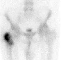
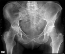
The bone
scan demonstrates abnormal tracer uptake in the right intertrochanteric region
typical for fracture (Figure 1). The corresponding pelvic radiograph showed no
fracture (Figure 2). The patient proceeded to internal fixation of the right
intertrochanteric fracture and rehabilitation.
Discussion
Hip fractures
are common and occur predominantly in women over 65 years. One year mortality
after this injury ranges from 15-20% and 50% of patients living independently
before sustaining a hip fracture are unable to regain their
independent
lifestyle. Delayed recognition can result in increased mortality
and morbidity. Symptoms are not always as typical as in this case and some
patients may have normal ambulation and
complain only of vague pain in their
buttocks, knees, thighs, groin or back. If radiographs are negative and symptoms
persist, or the clinical suspicion for fracture is high, a bone scan is an
appropriate next step. At 72 hours post injury, the
sensitivity of the bone
scan for detecting fracture is greater than 95%. A negative bone scan virtually
excludes fracture. Bone scans will detect a variety of other conditions which
may explain the patients’ symptoms. These include: fractures in other sites -
proximal femoral shaft, neck of femur, pelvis, sacrum and lumbosacral spine;
bursitis - ischial and trochanteric; enthesopathy; soft tissue inflammation;
metastatic disease; and arthritis - hip, facet and sacro-iliac joints. Following
falls, whole
body images are often performed to exclude fractures at distant
sites.
Conclusion
Bone scans are a highly sensitive appropriate investigation
to confirm or exclude suspected fracture following trauma in patients with
negative radiographs. In addition, alternate causes for the patients’ symptoms
can be detected and the whole body can be imaged. In suspected femoral fracture,
the early diagnosis and management is important to decrease morbidity and
mortality.
Reference: Brunner LC et al.
Hip Fractures in Adults. Am Fam Phys 2003: 67: 537-42
Case Study submitted by
Dr Scott
Beuzeville
Department of Nuclear Medicine,
St. George
Hospital.