What is a renal perfusion scan? The scan detects the presence of significant renal artery stenosis that is responsible for hypertension. The scan can also be used to detect urinary tract obstruction.
When should I order a renal perfusion scan? 1. Young patients with newly diagnosed hypertension for which no
other cause can be found
2. Patients with difficult to control hypertension
3. Patients with hypertension with small kidney on other imaging
4. Patients with hydronephrosis
What do I tell my patient?
Allow 2 hours.
Hypertensive patients.
Ideally, ACE inhibitors and Angiotensin II antagonists should be stopped for 48 hours before the scan. However this is not always possible. No other preparation is needed.
The patient should not fast. Upon arrival in the Department, the patient will be given a tablet of Captopril and be asked to drink several glasses of water over the next hour. The patient is then transferred to a scanning bed and given an intravenous injection of tracer and imaged continuously for thirty minutes. The patient is then free to go. The study is then analysed and if
the Captopril provoked study is abnormal, then a further scan is needed and the patient is contacted to arrange for a second scan.
Hydonephrotic patients.
Allow 2 hours.
No preparation required. The patient is given an intravenous injection of tracer and imaged continuously for thirty minutes. At the end of that time, the patient is given an injection of frusemide and a further thirty minutes of imaging is performed.
What will the scan tell me?
In hypertensive patients, the scan will detect significant renal artery stenosis that is responsible for the hypertension. In hydronephrotic patients, the scan will determine if the hydronephrosis is due to obstruction or not.
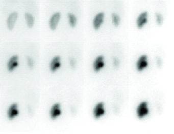
Tracer early and then the tracer washes out of the kidney. The kidney on the right progressively accumulates tracer. The patient is then administered frusemide intravenously with further imaging to estimate whether the kidney is obstructed
.What next?
Positive scans demonstrating renal artery stenosis usually require referral to a renal physician. Similarly, obstructive finding require referral to a urologist.
What is a renal cortical scan?
The scan detects the presence of renal scars in children.
When should I order a renal cortical scan?
1. Patients with vesicoureteric reflux
2. Patient with recurrent urinary tract infections
What do I tell my patient?
Allow 4 hours.
No preparation required.
The scan should not be performed for 10-12 weeks after a UTI.
The patient is given an intravenous injection of tracer and imaged 3 hours later. There are no restrictions on the patient during the three hour break.
What will the scan tell me?
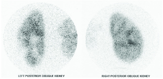
The scan clearly delineates the cortical outline of each kidney and renal scars can be visualised.
DMSA scan on four month old infant with a past history of urinary tract infection and vesicoureteric reflux. There is evidence of renal scars involving the lower half of the left kidney with a smaller scar in the posterior right kidney.
What next?
Positive scans demonstrating renal scars usually require referral to a renal physician.